


Lumbar osteochondrosis is a chronic degenerative-dystrophic disease of the lumbar spine, which affects the structure of the intervertebral discs and a number of lumbar vertebrae.This affects people at mostly working age.It manifests itself in various symptoms, the main of which are pain in the lower back and legs, limiting movements in the lower back.Studies such as radiography, computed tomography or magnetic resonance imaging of the lumbar spine are used for diagnosis.In this article, you can more detailed with the causes, symptoms and methods of diagnosing osteochondrosis of the lumbar spine.
Osteochondrosis is the result of body aging.These or other signs of this disease can be found in almost any person (!), From 25 years old.But here is the severity of these changes, the rate of their progression, the degree of clinical manifestations depends on many causes, primarily on how a healthy lifestyle leads a specific person.Moderate physical activity, mandatory morning gymnastics, proper body posture when performing a number of works (garden, construction, banal cleaning of the house, etc.), orthopedic mattress are those moments that prevent the development of lumbar spine osteochondrosis.
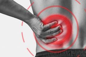
According to statistics, osteochondrosis of the spine in 80% of cases is the cause of back pain.
How does osteochondrosis develop?
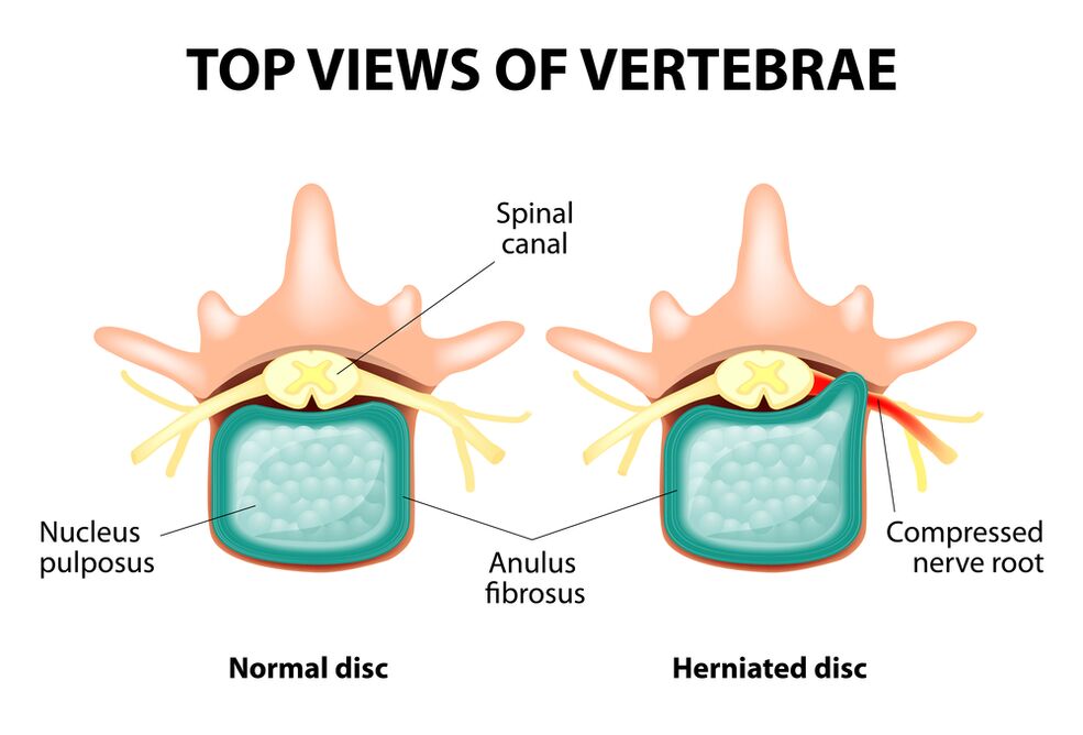
The entire spine consists of separate vertebrae, between the bodies of which there are intervertebral discs.That is, between the two vertebrae is one disc.The disc consists of a gelatin (pulpic) nucleus and a fibrous ring.The nucleus contains a lot of water and provides depreciation and flexibility of the spine.The fibrous ring is located along the periphery of the jacket, as if holding it inside.
With prolonged increased load on the core of the port, it changes its physiological properties, loses water and dries and ultimately sequences: the disc is flattened and the vertebrae bodies approach each other.Along with similar processes, in the kernel jacket, the fibrous ring loses its elasticity and, under the influence of mechanical loads, begins to protrude.This is called convexity.Then the fibrous ring cracks and the gelatin nucleus falls through the gaps obtained: the hernia occurs on the disc.A schedule of two adjacent vertebrae and a disk located between them, called the vertebra segment, acquires unnecessary mobility, thus increasing the load of nearby segments.The overload of adjacent segments triggers a similar pathological process in them.These changes are called osteochondrosis.
To ensure the stability of the spine in some way, bone growth is formed at the ends of the vertebrae bodies, increasing the support area.This phenomenon is called spondylosis.Changes in the joints between the vertebrae are called spondilo arthrosis.Usually all three pathologies - osteochondrosis, spondylosis, spondyl arthrosis - a walk nearby.
Reasons
Why does osteochondrosis occur?To date, there are several theories about the event:
- Mechanical theory: Perhaps the main reason should be considered a regular increase in spine.Therefore, osteochondrosis is an almost mandatory fate of hamals, miners, builders and people from such professions.The appearance of osteochondrosis of the lumbar region is mainly associated with slopes and lifting of weight, forced uncomfortable work posture;
- Another developmental factor is an incorrect posture sitting in the wrong posture, which is especially important for mental workers;
- Sometimes the role is played by hereditary characteristics of the structure of the spine and the nutrition of its individual structures;
- Traumatic theory: Any trauma to the spine (even the most miserable) is able to initiate a degenerative process;
- Hormonal metabolic disorders and endocrine diseases can adversely affect the metabolism in the tissues of the spine and contribute to the development of osteochondrosis;
- The theory of age implies the natural wear of the disks in the process of life.
Rarely only one of these theories can explain the appearance of osteochondrosis in any case.More often, at the same time, several factors are "guilty".
When osteochondrosis of the lumbar spine occurs, overweight plays an important role as it itself is overload for the spine.The higher the body mass index (degree of obesity), the more pronounced changes in the spine are usually.Among other causes provoking the onset of osteochondrosis, it may be noted:
- A sedentary lifestyle;
- Imorly Nutrition (fast food, unnecessarily sweet, midfield products: all this leads to an imbalance of trace elements) and lack of fluid;
- Abnormalities of the structure of the spine (for example, the presence of additional lumbar vertebrae);
- Constant wearing of high mill shoes;
- pregnancy (due to excess loading of the lumbar spine);
- sudden termination of training in people professionally involved in sports;
- Smoking and alcohol abuse: as factors that speed up the aging process in the body.
Symptoms
The main manifestation of osteochondrosis of the lumbar spine is the pain.The nature of the pain, the site of occurrence and the direction of distribution depend on which receptors are irritated, that is, how rough changes in the disc and surrounding tissues have a convexity or hernia, in which a convexity is formed, etc.
Reflex syndromes and compression are distinguished by osteochondrosis of the lumbar spine.
Reflex syndromes develop in cases where the receptors of the fibrous ring of the affected disc, ligaments and joint capsules located nearby are irritated.They are reflexive because in addition to the pain they are accompanied by muscle tonic, vegetative-vascular or neurodystrophic reflex changes, that is, irritation with reflexes is transmitted to other structures, causing symptoms mainly on the side of the soft tissues.
Compression syndromes occur as a result of compression (compression) of nerve roots, blood vessels or spinal cord formed by osteochondrosis by change.

Reflex syndromes of lumbar spine
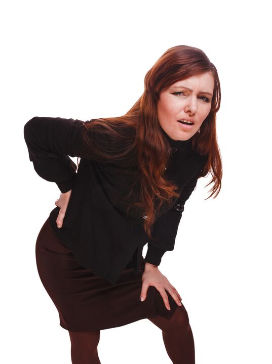
Lumbago(Feeling): Acute sudden pain in the lower back, which occurs with uncomfortable movement or during physical tension (much rarer - for no apparent reason).The appearance of Lumbago is believed to be related to the movement of the collar in the fibrous ring, that is, develops in the initial stages of osteochondrosis.Often the pain is described as "feeling", "the bet is stuck in the lower back."Patients freeze in the posture in which the pain has caught them.The smallest move causes an increase in pain (sneezing, coughing, trying to turn to bed, move your leg).If a person was in a sloping position during the development of Lumbago (which happens most often), then he cannot stand up.Muscle tension in the lumbar spine appears reflexively.Along the vertebrae in this area, there is a muscle roll, which is sometimes seen in the naked eye without touching and muscle tension is so pronounced.I feel painful for the patient.Such increased muscle tone plays an immobilizable role, protecting the affected lumbar segment of pathological mobility, which can cause worsening in the state.The natural bend of the spine in the lower back (lordosis) is flattened, perhaps curvature (scoliosis) is possible due to muscle tension.
Lumbalgia- Another reflex syndrome at lumbar level.This term also means the presence of pain in the lumbar region.But unlike Lumbago, the pain does not occur sharp, but gradually, within a few hours or even days.The pain is stupid, moderate intensity, increases during movements, in a sitting or standing position when moved from one position to another.A little relief is the position of a lying or the back with a roller under the lower back, but the passive elevation of the upright leg in this position causes increased pain in the lower back (symptom of lasa).The palpation of the lumbar spine is painful, but the reflex muscle tension is more pronounced than in lumbago, and sometimes it is absent at all.Movements in the lumbar spine are limited, but it is possible.This means that the patient can bend down and sideways to a certain level (and then the pain intensifies).
Sciatica- Another variety of reflex syndrome at the lumbar level.This term means pain in the lower back, which gives the butt and the legs (on the back surface).The pain is different, mostly ill, but can be increased periodically by the type of "fireplace" in the leg.Just as with Lumbalgia, it is enhanced by any movement, walking, tension, decreases when lying on the back.The symptom of lasa is usually positive.The palpation of the lumbar spine is painful, as well as the push of some points (for example, in the middle of the line that separates the ass from the thigh, in the middle of the back of the thigh, in the middle of the poppy pit).There is tension of the muscles of the lower back.The inclinations forward and on the side are limited.
Lumbar spine compression syndromes
The clinical characteristic depends on which structure is subject to compression.
Between the vertebrae in each intervertebral openings are nerve roots (spinal nerves): left and right.If the pathological formations for osteochondrosis of the lumbar spine (mainly discs on the discs) press the roots, then radiculopathy develops, the symptoms of which differ for each root.In total for all radiculopathies of the lumbar region is the increase in pain during sneezing, coughing, movement in the lower back (especially tilting forward), the presence of muscle tension in the lower back, limiting movements in the lumbar spine.The following types of radiculopathies of the lumbar spine are the most common:
- Radiculopathy L1, L2, L3: The pain occurs in the lower back, give the expected thigh.In the same area, paresthesia (a feeling of creeping geese, numbness) is possible, the surface sensitivity is disturbed (the sharp touch of the usual is not different, the feeling of cold and hot) is lost.Knee reflex decreases, the weakness of the quadriceps of the thigh is revealed;
- Radiculopathy L4: The pain from the lower back gives the front of the thigh, the inner surface of the knee joint and slightly lower along the inner surface of the lower leg.Paresthesia is felt in the same areas and surface sensitivity is lost (decreases).The weakness in the quadricema muscle of the thigh also develops, the knee reflex decreases;
- Radiculopathy L5: One of the frequent locations.The pain adds to the ass, along the outer edge of the thigh, along the anterior surface of the lower legs to the inner edge of the foot and thumb.Paresthesia is felt here, surface sensitivity is impaired and there is a sneeze and cough pain.In addition, there is a difficulty in extending the thumb of the foot, since the muscle that performs this action is innervated by the Kine L5.Sometimes it is difficult to stand on the heel with an open leg;
- S1 radiculopathy is also commonly found with osteochondrosis of the lumbar spine.The pain adds to the ass, along the outer edge of the thigh, along the outer edge of the lower leg to the outer edge of the foot and the 5th toe, the heels.These areas are characterized by a sense of paresthesia, reducing surface sensitivity.The Achilles reflex is reduced.With damage to this spine, the weakness of the muscles of the lower legs and flexors of the foot develops, so standing and walking on the socks is difficult.
The simultaneous development of radiculopathies of several roots is possible, this is especially characteristic of the L5, S1.It happens that a hernia squeezes several roots.
If the flock of the disc is blind, then it can pull out the spinal cord.This is only possible when the hernia is located at the upper reference point, since there are no spinal cerebral vertebrae below the lumbar vertebrae (the roots of the spinal cord are subjected to compression and the ponytail syndrome develops).
If the vessels of the lumbar region are subjected to squeezing, which perform blood flow to the spinal cord, then in the case of acute circulation, a spinal stroke develops and with prolonged compression - myelopathy.Myelopathy is manifested by bilateral weakness of the leg muscles, starting from the foot and gradually progresses upwards.The sensitivity in the legs is impaired, the Achilles reflex is lost, and later the knee.Urination disorders may occur (an honor, a "imperative" impulse requiring immediate satisfaction, urinary incontinence).
Diagnostic methods
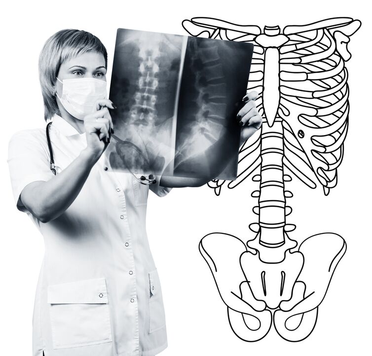
The diagnosis of osteochondrosis of the lumbar spine is based on clinical data and data for additional examination methods.The key role belongs to such methods such as:
- Radiography of the lumbar spine;
- Computed tomography of the lumbar spine;
- Magnetic resonance imaging of the lumbar spine.
The radiography of the lumbar spine is necessarily performed in 2 mutually perpendicular projections-the rights at the back and the side.Such photos allow you to see the shape, contours and structure of the vertebrae bodies, the height and shape of the intervertebral discs, the anomalies of the spine and the natural turns.In order to show the intervertebral joints and intervertebral openings, the radio grams are manufactured in inclined projections.In order to identify the pathological mobility of individual lumbar segments (which is a sign of osteochondrosis), radiography is performed under the conditions of functional testing, that is, in flexion and expansion of the spine.Usually you can clearly see the change in the height of the intervertebral discs in the front or back according to the direction of inclination of the body, with osteochondrosis due to the functional block of one of the segments, the height of the disc does not change either when bending or lengthening.Pathological mobility determines the displacement of the vertebrae forward or backward.The main X -branched signs of osteochondrosis include narrowing of the intervertebral percentage, pathological mobility and displacement of the bodies of the spine, deposition of salts in the disc tissue (calcification), the formation of regional outgrowths of the spinal bodies, the formation of the spine at the border (s) with the affected discRadiography of the lumbar spine is a routine of study that gradually loses its importance against the background of active application of new and more information methods of study (CT and MRI).The radiography of the lumbar department is today used as a screening diagnostic method.
The CT of the lumbar spine is also performed with the help of X -ray radiation, but the radial load on the body is much smaller than with X -Ray.The study is conducted, lying on the table of a special device - a computer tomograph, it is absolutely painless.The resulting photos are processed using a computer and allow you to see significantly more structures than with radiography of the spine.
MRI is a method in which electromagnetic radiation is used to create images.The study is also carried out in the position of lying on the table, which is called in the camera of the Tomograph.MRI is harmless and painless.
CT or NMR of the lumbar spine allow you to see all the structures of the spine, carefully examine the intervertebral discs (and the jacket and the fiber ring) and the intervertebral openings, the contents of the spinal canal.Even a slight convexity of the intervertebral disc will not go unnoticed.These methods (especially MRI) allow you to determine the direction of disc herniation, if any, the degree of compression of nerve roots, spinal cord.Thus, these methods of examination are much more information in the diagnosis of osteochondrosis of the lumbar spine than radiography.In addition, they allow you to diagnose not only osteochondrosis, but also other diseases (tumors, blood disorders in the spinal cord, abscesses, congenital defects in the structure of the spinal cord and spinal cord), which is important during the differential diagnosis of the causes of back pain.
Osteochondrosis of the lumbar spine is a disease that most often causes back pain.In fact, it is the destruction of the intervertebral discs.Due to osteochondrosis of the lumbar spine, a person often loses working capacity, since in addition to pain the disease can lead to impaired spinal motility, inability to sit, stand and walk.The symptoms of this disease are non -specific and require additional examination methods to confirm the diagnosis accurately.The most informative and safe for modern methods for diagnosing osteochondrosis is a spinal cord.