


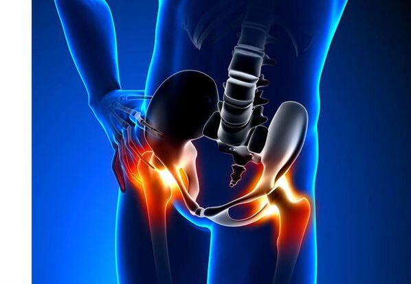
Hip Arthrosis (Coxarthrosis)- This is a chronic degenerative disease of the joints, which leads to deformity of bone tissue.With coxarthrosis, all components of the joint are involved in the pathological process: joint cartilage, bone structures adjacent to cartilage, synovial shells, ligaments, capsule and adjacent muscles.In the case of disease, articular cartilage is destroyed, micro-precipitation bones and osteophytes (bone growths) appear and inflammation of the musculoskeletal apparatus of the hip joint occurs.
In the world, every fifth person complains of joint problems with the joints.This can be both pain and movement restriction in the joints and a combination of these symptoms.Each second outpatient vision falls on patients with musculoskeletal disorders, while 66 % of cases are people under 65 years of age.According to recent epidemiological studies, the spread of arthrosis of the knee and hip joints among the adult population is 13 %.
Risk factors for the development of coxarthrosis:
- Genetic predisposition.A common cause of coxarthrosis of the hip joints is the congenital or acquired mutation of the type II prolagen type.
- Old age.The probable cause of the spread of arthrosis in the elderly is a discrepancy between the harmful effect on the joint cartilage of the external environment and its ability to recover.
- Under.Women suffer from osteoarthritis more often than men.This is due to the effects of the influence of female sex hormones of estrogen on bone-mineral metabolism.However, the influence of the floor is ambiguous - according to some authors, unlike the damage to other joints, there are no differences in the sexual basis for coxarthrosis: in men, hip arthrosis occurs as common as in women.
- Excess body weight.The connection has been proven between excess body weight and the onset of arthrosis.Excess adhesive tissue increases the harmful load on cartilage.In addition, adipose tissue produces pro -inflammatory enzymes that damage cartilage tissue.
- Frequent development of bones and joints.In accordance with studies, 80 % of coxarthrosis, which occurs for no apparent reason, is related to the fact that defects in the development of the hip joint - dysplasia and subluxation are not diagnosed.
- Hard physical labor.Excessive loading of the hip joints with certain types of physical labor can lead to damage to the joints and the formation of arthrosis.Agricultural workers, diggers and people with similar working specialties are at risk.
- Injuries.The risk of developing coxarthrosis increases after injury to the hip joint.In addition, one wounded joint may be involved in the process.
- Professional sport.Professional sports can provoke the appearance of coxarthrosis both due to excessive joint load and due to injuries.Potentially dangerous sports include heavy athletics, athletics jumping, parachute sports.
- Bones and joint diseases- Rheumatoid arthritis, psoriatic arthritis, joint infections, avascular necrosis, gout arthritis and more.
- Endocrine pathologies- hypothyroidism, hypoparathyroidism, acromegaly (impaired function of the anterior pituitary gland), diabetes, obesity.
If similar symptoms are found, consult a doctor.Don't be aware - it's dangerous to your health!
Symptoms of arthrosis of the hip joints
The main symptoms of coxarthrosis include: pain, restrictions on mobility and crunching in the joints, their deformity, functional shortening of the lower limb and periodic swelling in the joints.
Pain of varying intensity.The pain in the joint is initially insignificant and occurs in a short time.They appear or exacerbate while walking or with other physical exertion, such as during squats, inclinations and weight lifting.As the disease develops, the pain intensifies and even a long break does not bring relief.In addition, the pain occurs with prolonged immobility and the fixing of the joint in one position.
Patients complain of the so -called "start -up" pain in the hip joints after sleep, driving in a car and other prolonged immobility."Starting" pain for coxarthrosis lasts no more than 30 minutes.The pain is exacerbated during hypothermia or in a stressful situation.They can be located in the area of the ass or groin, on the anterior or lateral surface of the thigh.With the spread of pain on the nerves of the lumbar plexus, it can be transmitted to the hips away from the center of the body or in the knee.Sometimes the pain is applied to the lumbosacral spine and tail.
Restricting joint mobility.Movements in the hip joint with coxarthrosis are limited due to pain.At the same time, rotation (rotates both inside and outside) and bringing the lower limb (the movement in the middle of the body) is more often disturbed, but it can be limited (movement from the middle axis of the body), as well as flexion and extension.The inability to perform passive movements in the joint due to pronounced pain syndrome causes compensatory pelvic bias.The patient's gait changes, the buttocks are protruding back, the body deviates forward as it transfers weight to the damaged side.With bilateral impairment in patients with coxarthrosis, a "pathetic gait" is formed.
Coxarthrosis occurs periodicallyswelling in the jointwhich can be invisible due to the muscle and fat layers.Also the disease is characteristicCrystals in the joints during movement, their gradual deformation and functional shortening of the lower limb.
Often one joint is affected by the disease, then the process is applied to others.But sometimes arthrosis affects several joints at a time and poliosoarthritis occurs.Policteoarthrosis is characteristic of the elderly or with hereditary predisposition and concomitant diseases - bone, joint diseases and endocrine disorders.
Pathogenesis of arthrosis of the hip joints
In the pathogenesis of hip arthrosis, an important role is played through mechanical damage (injuries and microtraumas due to increased physical activity on the joint) and genetic, hormonal and metabolic factors.It is often not possible to understand which factor has influenced the development of the disease in a particular patient, but often the disease develops after damage to the tissues with mechanical injury.
Tissue damage stimulates the separation of cartilage tissue cells (chondrocytes), while the production of pro -inflaming cytokines increases, which are usually present in cartilage only in small quantities.Cytokines begin the inflammatory process, for example, under the influence of the pro-inflammatory cytokine IL-1, the enzymes that destroy the cartilage of the joint are distinguished.Also, under the influence of cytokines, the production of the tsog-2 enzyme and other substances that have a toxic effect on the increase in cartilage.
Synovites also play a big role in the development of coxarthrosis - inflammatory diseases of the synovial lining of the joints or connections with the accumulation of fluid in the cavity.
Reducing the elasticity and strength of articular cartilage associated with metabolic disorders leads to a decrease in its resistance to mechanical stress.With coxarthrosis, all joint components are involved in the pathological process, including subchondral bone.Due to the fact that large joints of the lower limbs take into account large joints of the body, they experience significant mechanical tension, which is why in the subchondral plate and cartilage and cartilage.As a result of the microvolomas, the subchondral bone is compacted, leading to the regional growth of bone tissue - osteophytes.And this, in turn, stimulates the larger degradation of the articular cartilage.
In some cases, hip arthrosis is inherited.Hereditary arthrosis is a polygenic heritage - due to the action of many genes, each of which influences poorly.The cause of some diseases is a mutation in genes encoding macromolecules of the articular cartilage, which causes its tears.The genes responsible for the division of chondrocytes can also suffer.In addition, metabolic disorders are inherited as pyrophosphate arthropathy - a disease in which the crystals of calcium pyrophosphate accumulate in the articular cartilage and synovial fluid.
Classification and stages of development of arthrosis of the hip joints
Depending on the causes of the disease, coxarthrosis is divided into two main forms: primary (idiopathic) and secondary (resulting from or due to other diseases).
Primary coxarthrosis:
- Localized (only hip joints):
- unilateral;
- bilateral.
- Generalized (polioaarthrosis) with a lesion of at least three joint groups (eg hip joint, knee and small joints of brushes or legs).
Secondary arthrosis:
- Post -traumatic:
- acute - as a consequence of acute injury;
- Chronic - due to classes of any sport or as a result of professional activity.
- Metabolic diseases (abnormal, hemochromatosis, Wilson's disease, Gauche's disease).
- Congenital pathologies and developmental defects (congenital hip dysplasia, affects the disease, slipping of the epiphysis of the femur, hypermobility syndrome, shortening of the lower limb, scoliosis, bone dysplasia).
- Endocrine pathologies (acromegaly, hypothyroidism, diabetes mellitus, hyperparathyroidism, obesity).
- Calcium salts (pyrophosphate arthropathy, calculating tendonitis).
- Bone and joint diseases (rheumatoid arthritis, psoriatic arthritis, pedetic disease, avascular necrosis, infections).
According to clinical manifestations, the following forms of coxarthrosis are distinguished:
- Little symptoms.
- A manifesto manifested by bright clinical symptoms:
- rapidly progressive, in which the symptoms develop during the first four years of the onset of the disease;
- Slowly progressive - clinically significant symptoms occur after five years of the disease.
In accordance with the photo X -Ray, two types of hip arthrosis can be identified:
- Hypertrophic - with signs of increased reparative response (lesions are replaced by new tissue, for example, osteophytes appear);
- Atrophic (reduction of tissue volume).
The stages of the disease can be determined radiologically and clinically.To determine the radiological stage of hip arthrosis, the classification of Kelgren and Lawrence (1957) is most commonly used.
Stages of arthrosis in radiological classification
| Stage | Signs |
|---|---|
| 0 | There are no signs of arthrosis in x -ray images |
| 1 | The joint difference does not change, single regional osteophytes are visualized |
| 2 | The joint difference does not change, significant regional osteophytes are visualized |
| 3 | The height of the joint gap is moderately reduced, significant regional osteophytes are visualized |
| 4 | The height of the gap of the joints is significantly reduced, significant regional osteophytes and subchondral osteosclerosis are visualized (sealing of bone tissue under the lower surface of the cartilage with the structure of the cartilage) |
A classification (1961) is used to determine the clinical stage of the disease, which uses both clinical signs and visualization criteria.
Clinical stages of arthrosis
| Stage | Signs |
|---|---|
| 0 | The articular precipice is narrowed unequivocally and unevenly, the edges of the joints are slightly directed (initial osteophytes), a slight restriction on movements is noted |
| 1 | The articular gap is significantly narrowed (50-60 %), significant osteophytes, subchondral osteocosclerosis and cystic enlightenment in bone epiphins;The clinic is prevalent from limiting mobility in the joints, rough crispy during movements, insignificant or moderate muscle atrophy |
| 2 | Deformation, stiffness of the joint;The articular gap is narrowed by more than 60-70 % of the norm or completely absent, extensive osteophytes, subchondral cysts, articular "mice" are visualized bone, cartilage or mixed pathological formations located in the joint cavity |
Complications of arthrosis of the hip joints
In coxarthrosis, all complications are related to the pathological changes in the joints.
The course of coxarthrosis can be complicated by local inflammatory processes:
- Bursite - inflammation of synovial bags in the joints;
- Tendovaginitis - inflammation of the inner sheath of the vagina of the muscle tendons;
- Pandom of the nerve tunnel syndrome due to the formation of large osteophytes or with joint deformity.
With the progression of coxarthrosis and its transition to the clinical stage II and III, the pain limits the mobility of the joint and over time appears the joint ankylosis (fibrous, bone or cartilage), accompanied by its complete immobility.
Significant joint deformation can lead toFractures or aseptic bone necrosis.For coxarthrosis, aseptic necrosis of the femur head is the worst complication.
With a pronounced coxarthrosis may occurSubluxation and dislocation of the jointas well as penetration of the femoral head into the pelvic cavity.The dislocations and subluxation of the hip joint lead to pain (initially acute, then dull and pain) that increase during walking and other physical activity, as well as to deformation of the joint, lame and sometimes to shorten the affected limb.
Despite the lack of systemic manifestations of arthrosis itself, more attention is paid to the diseases associated with it in modern clinical practice.These are such pathological conditions that exist or arise against the background of the present disease.In connection with inflammatory reactions occurring during arthrosis, the formation of atherosclerotic plaques on the internal walls of the vessels is enhanced, which increases the riskCardiovascular diseaseSReducing physical activity due to pain and restriction of joint mobility leads toobesity, depression and deterioration of quality of lifeSWith prolonged use of non -steroidal anti -inflammatory drugs,Are affected by the upper gastrointestinal sections,And tooThe risk of cardiovascular pathologies and kidney diseases increasesS
Diagnosis of hip arthrosis
The diagnosis of "coxarthrosis" is made on the basis of clinical manifestations and radiological examination.There are no characteristic laboratory signs for the diagnosis of arthrosis.
Among the clinical manifestationsThe main for the diagnosis of hip arthrosis is the pain and its character.The pain for arthrosis of the hip joint occurs and grows gradually for several years (sometimes several months with a rapidly progressive form).The pain occurs or increases during exercise or in an upright position.If the patient begins to experience pain alone, then the inflammation (synovitis) joins.A statement is noted within 30 minutes in the morning and with prolonged immobility.
The restriction of joint mobility is gradually increasing, this applies to both active and passive movements.With the development of the disease, the joints deform, a functional shortening of the length of the limbs may occur.
In studying physicsThere is a restriction on the mobility of the joints, their deformity, shortening of the limbs, pain in palpation of the joint and large rotation of the femur, muscle atrophy.
LaboratoryNo required to diagnose arthrosis of the hip joints.However, they can be used for differential diagnosis of coxarthrosis with arthritis (rheumatoid and chronic), since with arthrosis there are no inflammatory changes in the general blood test and rheumatoid factor and uric acid levels do not increase.In addition, using laboratory tests, contraindications for the methods of drug treatment are revealed.
Instrumental methodsTo diagnose arthrosis of the hip joints:
- Radiography- This is the main method of diagnosing arthrosis of the hip joints.Radiography determines the changes characteristic of coxarthrosis: narrowing of the joint precipice, osteophyte, erosion and ulceration of cartilage, subchondral cysts and osteosclerosis.X -ray study is a classic method for diagnosing coxarthrosis, and radiological features are at the heart of the classification of coxarthrosis.However, other methods for visualization of the joint, such as ultrasound and magnetic resonance, are currently increasingly used.
- Ultrasound (ultrasound) -The advantage of ultrasound is in the absence of radial load on the body.
- Magnetic resonance imaging (MRI)- Compared to other methods, it allows you to more clearly visualize the damage to the joints.
- Arthroscopy-Allows you to identify damage to the articular cartilage: from the zones of chondromation (softening of the articular cartilage) with a diameter of 10 mm to deep cracks that penetrate to the subchondral bone and the formation of deep ulcers.Surface and medium cracks and surface erosion can also be visualized.
Identification of coxarthrosis is usually not a special difficulty, but when assessing a specific clinical situation, it is necessary to remember the possible secondary origin of hip arthrosis (such as complications of other diseases, for example, with endocrine disorders).
Treatment of hip arthrosis
Treatment of arthrosis of the hip joints can be either conservative (medicine and not united) or surgically.Conservative treatment is used in 1-2 stages of the disease, the surgical stage in 3 stages.Surgical treatment can be recommended in 2 stages with constant pain and lack of reaction to conservative therapy.
The goals of conservative therapy:
- Improving quality of life - reduce pain and increase joint mobility;
- Stop or delay the development of the disease.
Treatment methods without medicine include:
- unloading of the hip joint (weight loss, creating additional support and transfer of part of the body weight in reeds or crutches);
- Physiotherapy Physical Education;
- Methods of physiotherapy treatment.
The treatment of coxarthrosis begins with methods that are not impaired, it gives an important role to physiotherapy exercises.In severe pain, the patient should use support.With a pronounced disease and the presence of contraindications to endoprotetics, support should be used for life.
Healing therapy of caorrosisIncludes medicines that reduce the symptoms of the disease.These are analgesics, as well as drugs in the group of non -steroidal anti -inflammatory drugs (NSAIDs).NSAIDs are divided into non -election and selective.
Analgesics and NSAIDs for hip arthrosis are used for a short time to relieve pain and inflammation.There is currently no proven advantage of one non -steroidal anti -inflammatory agent over another, so the choice of a particular drug depends on the side effects and a specific clinical situation caused by it.
It should be remembered that NSAIDs have a number of side effects.When you take them, the mucous membrane of the stomach and duodenum is affected, resulting in ulcers and bleeding.A number of NSAIDs have a toxic effect on the liver and kidneys.In addition, NSAIDs disrupt platelet aggregation and, as a result, the patient is disturbed by thrombosis and tends to bleed.NSAIDs with prolonged use suppress the processes of hematopoiesis and can cause aplastic anemia and agranulocytosis.The adoption of selective NSAIDs causes significantly less complications.
The iron and gels used topically cause less side effects than oral products.For the treatment of arthrosis, medicines with warming and pain reduction are used.They may contain turpentine, menthol, nicotinic acid, salicylates, bee venom.Also, NSAIDs have good effect.
In the absence of the effect of analgesic and NSAIDs, or if it is impossible to choose the optimal dose of the drug, the painkillers from central actions may be prescribed short -term.
In the case of inflammation, internal -articular administration of corticosteroids is used.Corticosteroids are used no more than 2-3 times a year, as more common use can lead to cartilage degeneration.
Slow -acting drugs weaken the symptoms of the disease include chondroprotectors, inappropriate compounds of avocado or soy, hyaluronic acid.These drugs are included in the recommendations of the European Anti -Rechematic League for the treatment of hip arthrosis.Preparations reduce pain and improve joint mobility.
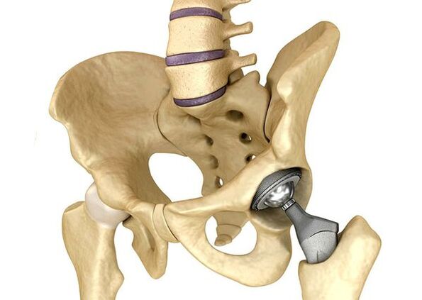
Hip endoprotheticsIt is used in severe cases of stage III when the pain syndrome cannot be eliminated and joint mobility is significantly limited.Hip prosthetics lead to a decrease in pain syndrome, improvement in the functional condition of the joint and the quality of life of the patient.The effect lasts 10-15 years, after which a second surgery may be required.During the operation, the hip joint is replaced by artificial imitation of ceramics, metal (most commonly used titanium dentures) or polymer.
Forecast.Prevention
The prognosis of arthrosis of the hip joints in connection with the life of the patient is favorable, but the disease often leads to damage.According to the World Health Organization, 80 % of adult coxarthrosis patients have a mobility disorder and 25 % cannot do daily questions.In this regard, the primary prevention of hip arthrosis is important.
Prevention measures:
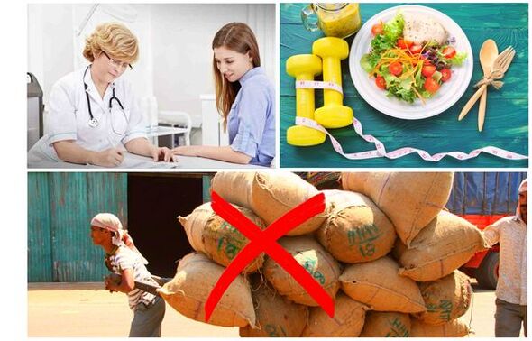
- Reduce body weightSIt is necessary to adjust the diet to reduce weight and load on the joint.In addition, reducing the volume of adipose tissue reduces the amount of mediators of inflammation it releases.
- Avoid heavy physical labor and sports overloads.Physical overloads are often the cause of hip arthrosis, while moderate physical activity, on the contrary, improves the condition of the articular cartilage, retains its normal mobility and reduces the load on other joints.
- Correct the underlying disease.If the patient is detected in diseases that can lead to secondary coxarthrosis (endocrine, rheumatic and others), the underlying disease is required.The normalization of hormonal origin and the achievement of a persistent remission of rheumatic diseases is as the main prevention of arthrosis, and allows you to slow down its development.
- Have a healthy lifestyle.A balanced diet with sufficient content of plant and animal proteins, polyunsaturated fatty acids and limiting simple carbohydrates, as well as moderate physical activity, avoid the appearance of coxarthrosis even in the presence of risk factors.
Currently, the prevention of hip disease is required in neonatology and pediatrics.Over time, the corrected congenital hip dysplasia significantly reduces the risk of coxarthrosis in adulthood.